Intraoral scans


These days, it’s a common occurrence for dental technicians to receive virtual impressions— digital scans taken directly in the mouth with a compact, ergonomic scanner.
But does this approach give accurate, reliable and, most importantly, usable results when it comes to producing a high-quality prosthesis? Without question, the short answer is yes. But could a dentist do the scan with their eyes closed? That’s another story.
The inside of the mouth is a tricky place to scan. It’s a wet environment with lots of different tissue types that are far from being completely opaque, which has a significant impact on the waves that are meant to “map” the site for restoration. The actual intraoral scans below demonstrate the current state of this technology. Let’s take a look at them.
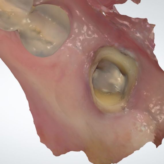
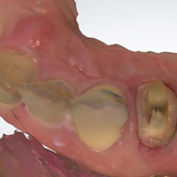
The latest generation of scanners can capture and display data in colour, for improved visualization of the different tissue types present.
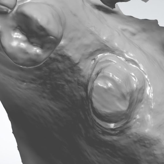
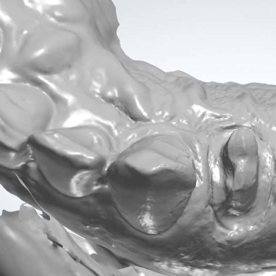
Here’s the same image in black and white. The monochrome option provides sharper images of the various textures in the mouth.
This new image seems to show rough patches on the distal surface. If we took the colour out of the equation, this area might be better defined.
In fact, this appears to be the case. The distal proximal contact is also clearer. If we look at tooth 37 on the previous colour image, we can see that scanners often produce a particular interpretation of metal fillings. Now we need to determine if the irregularities seen on the distal margin are actually part of the tooth or an anomaly that we need to work around.
Sometimes the opposite happens and the black and white image doesn’t clearly show a distinction between the soft and hard tissues.

Whereas the colour image shows it more clearly.
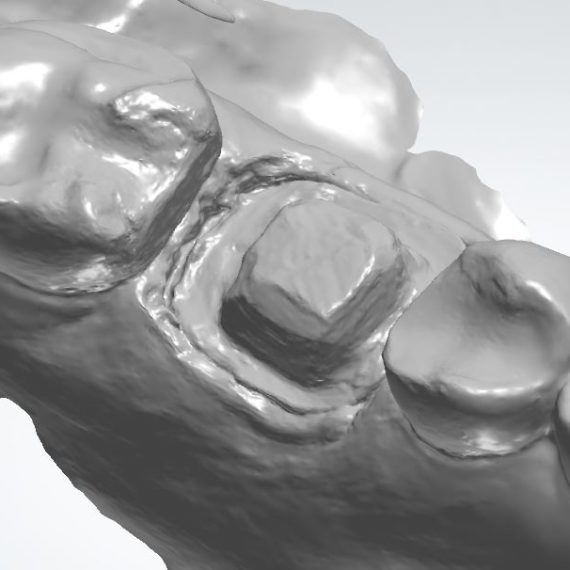
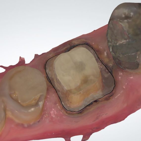
Some practitioners make more extensive use of the many options available in intraoral scanner systems. Here, the dentist drew the marginal finish line (black line) chairside, which limits the amount of interpretation the technician will have to do.
Here’s the same image but this time in black and white.
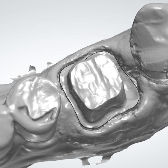
Since it’s a question of interpretation, certain areas sometimes need to be examined more closely by the technician. Here, the buccal finishing of the preparation seems to be clear at first glance.
But the monochrome image shows mucosa or some sort of fluid blocking the view of a small portion of the margin in question. A dental lab accustomed to using digital technologies will be able to identify insidious problems hidden in the raw data.
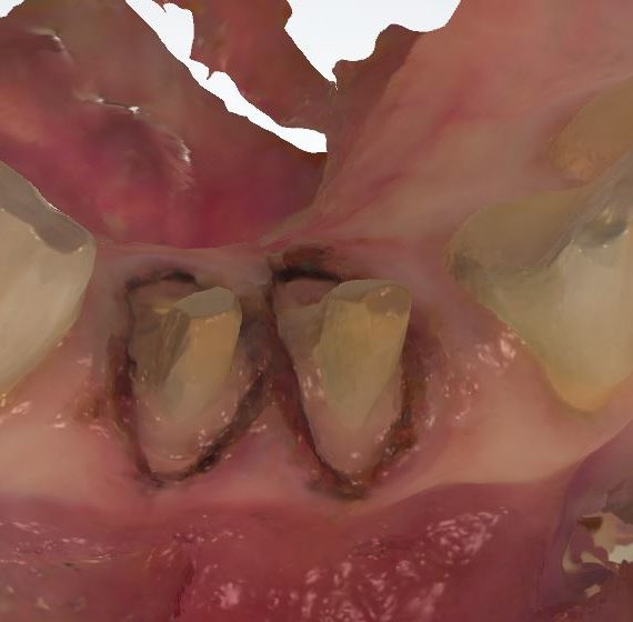
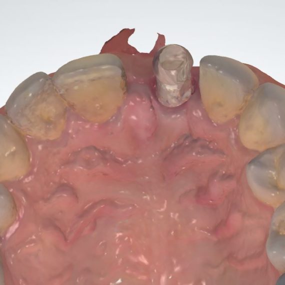
Restorations with implants can also be made using a direct scan, albeit with a few more steps. Here we can see the scan body in position 21.
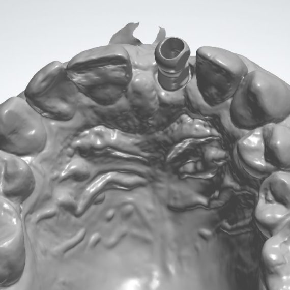
We see it better in black and white.
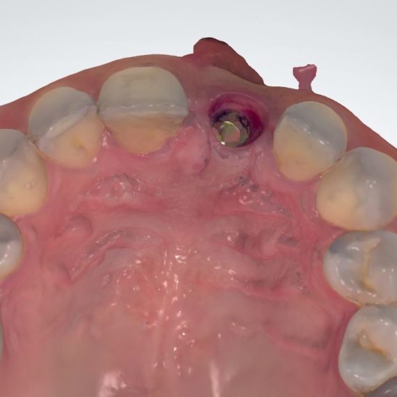
Don’t forget to scan the implant site without the scan body, so as to give the technician a better idea of what the mucosa around the implant actually looks like.
With a little effort and eyes wide open, the learning curve with this technology is quite manageable. And while it’s not a magic solution, it’s fast becoming an essential compo